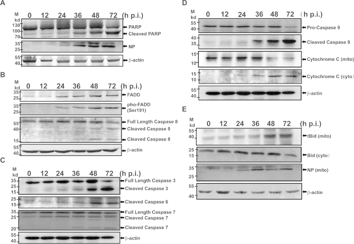
Figure 4. Apoptosis was activated in infected HepG2 cells.
Cell lysates were prepared from infected and uninfected cells at indicated time points from 12–72 hrs p.i., and resolved with 10–15% SDS-PAGE. Proteins were transferred to PVDF membranes for western blot analyses with specific antibodies. (A) PARP was cleaved in infected cells. (B) Upregulation and increased phosphorylation of FADD and increased cleavage and activation of pro-caspase 8. (C) Cleavage and activation of executioner pro-caspase −3,−6 and −7. (D) Cleavage and activation of pro-caspase −9 and release of cytochrome c from the mitochondrial membrane to the cytosol. (E) Cleavage and activation of Bid and translocation of tBid into the mitochondrial membrane.

