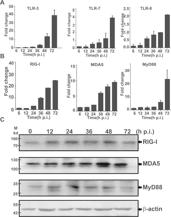
Figure 7. Induction of TLRs and RLRs in SFTSV-infected HepG2 cells.
Total RNA was prepared from SFTSV-infected or uninfected cells at different time points and used for reverse transcription and real-time PCR analysis with specific primers to respective genes. Reactions were performed in duplicates, each reaction was repeated for at least three times, and a representative result is presented. Fold changes were calculated relative to a gene in uninfected and infected cells, which were normalized to GAPDH. Fold changes in transcript levels for TLR-3, TLR-7 and TLR-8 (A); and MyD88, RIG-I and MDA5 (B). Cell lysates were prepared and subjected to SDS-PAGE and western blot analyses with antibodies against RIG-I, MDA-5, MyD88, and β-actin at the time points as indicated.

