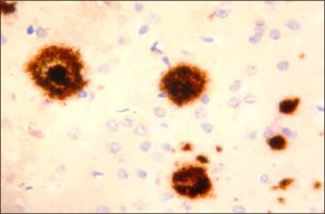
Fig 2.
An immunohistochemical section taken through the cortex in a case of ADD. An antibody to Beta A4 amyloid is applied to the tissue and detects this antigen which in turn stains the antigen brown. This shows a dense deposition of amyloid throughout the cortex as dense core (DC) and diffuse (D) plaques.

