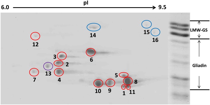
Fig 3. Identification of the gliadin protein spots from T. urartu after resolution with 2-DE.
Gliadins were prepared from mature grains, separated by 2-DE, and further identified via MALDI-TOF/TOF-MS analysis. Shown on the right side is the SDS-PAGE separation of prolamins from T. urartu. The high-molecular-weight glutenin subunit protein spots are not shown because of limited space. The spots in red circles are gliadins, the spots in blue circles are LMW-GSs (spot 14, KM085281, MW: 38.06, pI: 7.91; spot 15, KM085304, MW: 38.56 pI: 8.5; spot 16, KM085275, MW: 37.52, pI: 8.71), and the spot in purple is avenin-3 (TRIUR3_09156, MW: 35.1, pI: 7.66).

