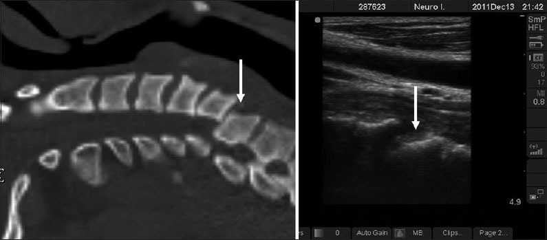
Figure 4.

Computed tomography cervical spine (sagittal section) and ultrasound image of the cervical spine of a patient with bilateral facet dislocation at C5–C6. The dislocation and disruption of the anterior longitudinal ligament is the cervical spine, is very well seen on ultrasound imaging (arrow)
