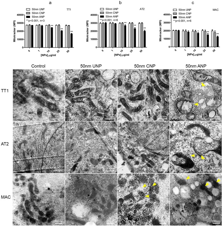
Fig. 4.

Effect of 50 nm polystyrene nanoparticles on mitochondrial membrane potential (a-c) and structure (d-o) following 4 h exposure. ANPs caused a significant reduction in the mitochondrial membrane potential (**p < 0.001, n = 3 TT1 replicates and 6 subject samples for AT2 and MAC) of all cell types (a-c) and altered mitochondrial structures by causing mitochondrial swelling (arrows in g, k, o) compared with the control (d, h, l). There was only slight mitochondrial swelling in MACs following CNP exposure (arrows n) not seen following UNP exposure (m). The number of total observed cells analysed/sample was 60 (n = 60); scale bar 500nm
