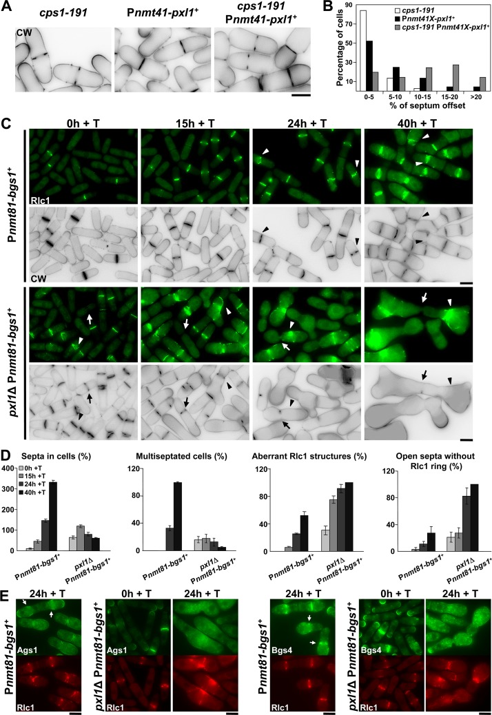
Fig 3. Cooperation of Bgs1 and Pxl1 is essential for CAR maintenance and septum formation.
(A) CW staining images of cps1-191, Pnmt41-pxl1 + and cps1-191 Pnmt41-pxl1 + cells. Cells were grown at 25°C (permissive temperature for cps1-191) in the presence of thiamine (+T, pxl1 + repressed) for 24 h and imaged. (B) Histogram showing the indicated intervals of positions of the septa measured as the percent of septum offset from the cell center: white bars, cps1-191 cells (n = 37); black bars, Pnmt41-pxl1 + cells (n = 44 cells); and grey bars, cps1-191 Pnmt41-pxl1 + cells (n = 62 cells). The percentages of septum offset were calculated as described for the Fig 1B. (C) Fluorescence micrographs of Pnmt81-bgs1 + and pxl1Δ Pnmt81-bgs1 + cells stained with CW and carrying Rlc1-GFP. Cells were grown to early log-phase in EMM+S (time 0 h), shifted to EMM+S+T for bgs1 + repression (times 15, 24 and 40 h + T), and imaged at the indicated times. Arrow: Cell with an open septum without the ring of Rlc1. Arrowhead: Cell with an aberrant ring of Rlc1. (D) Histograms showing the indicated percentages of septa and Rlc1 structures in Pnmt81-bgs1 + (n = 150 cells or hypha units for each time), and pxl1Δ Pnmt81-bgs1 + cells (n = 100 cells or hypha units for each time). Note that Pnmt81-bgs1 + strains at 24 h of bgs1 + repression appear as hypha units, each being equivalent to several single cells. (E) Fluorescence micrographs of Pnmt81-bgs1 + (time 24 h of bgs1 + repression) and pxl1Δ Pnmt81-bgs1 + (times 0 and 24 h of bgs1 + repression) cells carrying Rlc1-RFP and Ags1-GFP or GFP-Bgs4. Cells were grown as in C and imaged at the indicated times. Arrow: concentration of Ags1-GFP or GFP-Bgs4 in the septum membrane. Scale bars, 5 μm.

