
Fig. 9.
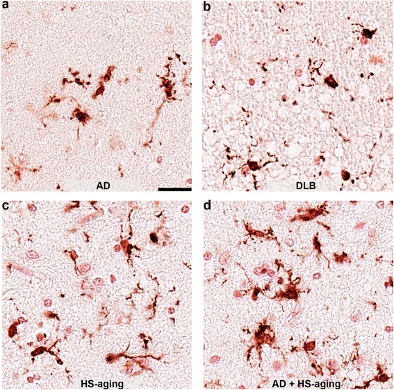
Dystrophic IBA1+ microglia in the hippocampus. Examples of IBA1+ dystrophic microglia in the CA1 region of AD individual (a; case #20), DLB individual (b; case #33), HS-aging individual (c; case #15), and AD + HS-aging individual (d; case #27). Scale bar is 25 μm