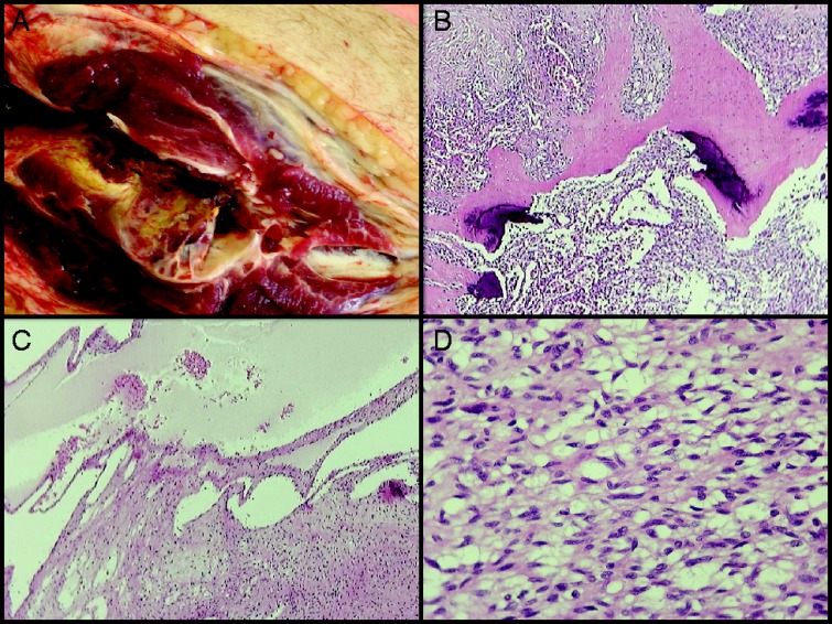
Figure 3.

Histology report. Dissection of the specimen showed multiple soft tissue tumors in all muscle compartments of the calf, measuring up to 18 cm (A). A separate lesion was found in the distal tibial metaphysis. Histological examination of all lesions showed a cellular spindle cell neoplasm with variously sized vessels, vascular-like spaces (B) and scattered deposits of calcified extracellular matrix (C). The neoplastic cells were arranged in bundles or diffusely. One small site was found to be highly pleomorphic and with high mitotic index (including some atypical mitosis) was found. The tumor infiltrated skeletal muscles, subcutaneous fat and the proximal end of the fibula. The immunohistochemical examination of the neoplasm showed positivity for vimentin while the markers for SMA, caldesmon, desmin, S-100 protein, EMA and CD34 antigen were negative. The cellular proliferation marker Ki67/MIB1 was positive in <10% of the neoplastic cells. The tibial lesion had identical histology and was separate from all other lesions. There were no signs of DLBCL infiltration. (A) The larger tumor in the upper calf, in close proximity to the proximal end of the fibula infiltrating the adjutant muscle compartments and subcutaneous tissue. (B) H&E stain ×20. Spindle cell neoplasm with both solid pattern and cystic, ‘vascular-like’, spaces. (C) H&E stain ×40. Scattered deposits of calcified extracellular matrix. (D) H&E stain ×400. Cellular spindle cell neoplasm with ‘benign’ histopathological features.

 This work is licensed under a