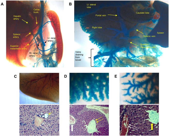
Fig 1. Selective angiography using blue latex dye.
(A) Systemic arteriography and (B) direct portography in a wild-type mouse. The stomach, duodenum, and jejunum were removed to allow a clear view of the vessels. (C-E) Magnified images of the liver after selective angiography and subsequent tissue clearing procedures (upper panels) and histological assessment (lower panels, hematoxylin-eosin staining) were performed. (C) Arteriography, (D) portography, and (E) hepatic venography specimens are shown. Blue latex dye particles are seen only in the target vascular compartment (yellow arrows), which seems to have been slightly dilated during injection (single arrow, hepatic artery; double arrow, portal vein; thick arrow, central vein). Blue latex dye particles are not detectable in non-target vascular compartments (white arrows).

