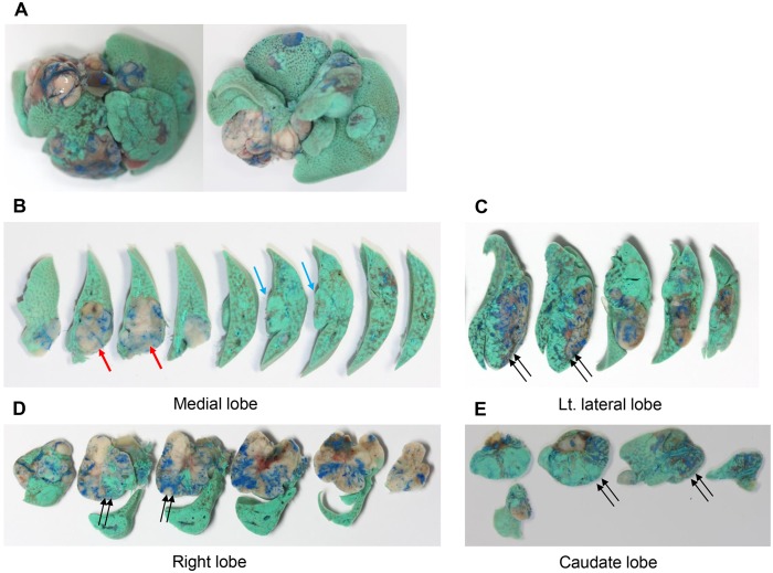
Fig 2. Liver dual angiography performed in a H-ras12V over-expressing transgenic mouse that developed multiple spontaneous tumors.
Selective latex arteriography (blue latex) and portography (green acrylic paint) were sequentially conducted. Gross images of (A) the whole liver superior aspect (left) and inferior aspect (right), and sectioned slices of (B) the medial lobe, (C) left lateral lobe, (D) right lobe, and (E) caudate lobe. Nodules were perfused by arterial supply (red arrow), portal venous supply (blue arrow), or both (double arrows).

