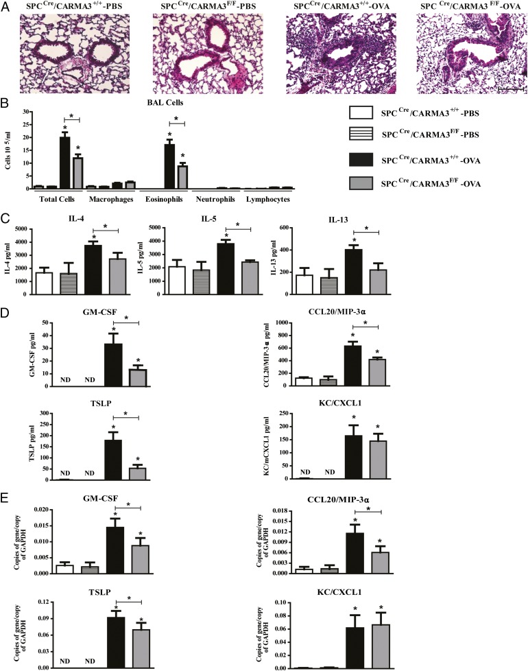
FIGURE 4.
Attenuation of airway inflammation in OVA-immunized and -challenged SPCCre/CARMA3F/F mice. (A) Histopathologic analysis of lung sections stained with H&E from SPCCre/CARMA3+/+-PBS–, SPCCre/CARMA3F/F-PBS–, SPCCre/CARMA3+/+-OVA–, and SPCCre/CARMA3F/F-OVA–treated mice. Scale bar, 50 μm. (B) Total cells, macrophages, eosinophils, neutrophils, and lymphocytes were enumerated in BAL fluid. (C) Protein levels of IL-4, IL-5, and IL-13 in the lung quantified by ELISA. (D) Protein levels of GM-CSF, CCL20/MIP-3α, TSLP, and CXCL1/KC in the BAL quantified by ELISA. (E) RNA levels of GM-CSF, CCL20/MIP-3α, TSLP, and CXCL1/KC in the lung quantified by qPCR. Data are means ± SEM of eight mice per group from two experiments. *p < 0.05 (OVA treatment compared with same genotype that was treated with PBS or SPCCre/CARMA3+/+ OVA compared with SPCCre/CARMA3F/F OVA). ND, not detected.

