
Figure 2.
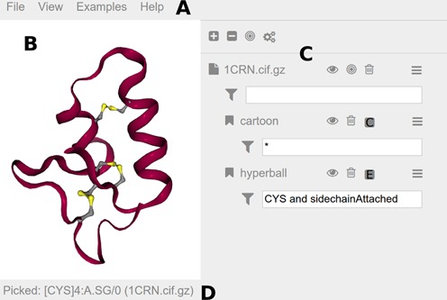
Screenshot of the ‘NGL Viewer’ GUI, magnified for clarity. A more detailed description of the GUI can be found in the online documentation. (A) The menu bar at the top provides access to general commands. The ‘File’ menu includes commands to load files or export images. Buttons to change the theme or to go fullscreen are in the ‘View’ menu. The ‘Examples’ menu includes various possible use cases. Help-related items like a preferences section and a link to the documentation can be found in the ‘Help’ menu. (B) Molecular visualizations are rendered to the viewport at the center. Here the structure of ‘crambin’ (PDB entry 1CRN) is shown highlighting the three disulfide bridges. (C) The sidebar hosts an interface element for each loaded structure and added representations. A number of buttons are available to quickly access commands: hide/show (eye icon), center (bulls eye icon), delete (trash bin icon) and parameters menu (stacked bars icon). Input fields for atom selections (funnel icon) restrict the display of representations. Here the ‘hyperball’ representation is limited to CYS and sidechainAttached. (D) The status bar at the bottom notes the last picked atom, in this case the ‘SG’ atom in the ‘cysteine’ with residue number ‘4’ in chain ‘A’ of model ‘0’.