
Figure 3.
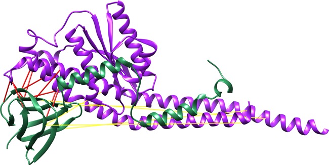
Top 15 interprotein scores overlapped between the four methods. Structure of the E. coli F1-ATP synthase (pdb: 3OAA), chain H (green) and G (violet). The distance of the top scoring pairs is depicted with red lines (Cα distance < 12 Å) and yellow lines (otherwise).