
Figure 2.
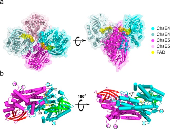
Overall atomic picture of ChsE4-ChsE5. (a) The biological functional unit is an α2β2 heterotetramer with two ChsE4 chains and two ChsE5 chains. The tetramer has two flavin adenine dinucleotide (FAD) binding sites, and two FADs are bound. The surface representation of the structure corresponds to the chain color. (b) A ChsE4-ChsE5 αβ heterodimer is shown as a cylindrical cartoon and colored by the secondary structure. FAD binds to the interface of ChsE4 and ChsE5. The structure on the right is a 180° rotation around the X axis of the structure on the left.