
Figure 1. Identification of an Intermediate stage of Yersinia Entry into Host Cells.
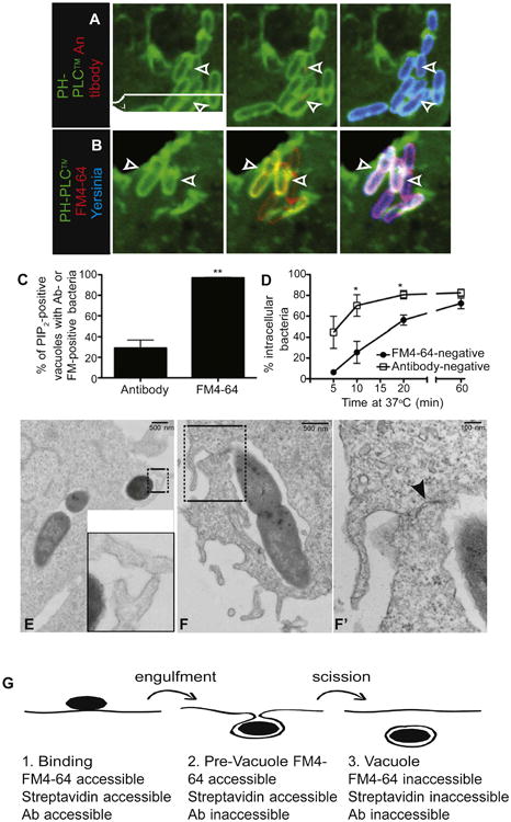
(A) Prior to vacuolar scission, Yersinia reside in PI(4,5)P2-positive structures that are inaccessible to antibodies. COS cells expressing the PH domain of PLCδ fused to GFP (PH-PLCδ) were infected with Alexa 647-labeled Y. pseudotuberculosis (blue) for 10 min, followed by fixation and staining using antibody to Yersinia, followed by Alexa 546-conjugated secondary antibody (red). Arrowheads denote PI(4,5)P2-positive vacuoles containing antibody-negative bacteria.
(B) PI(4,5)P2-positive, Yersinia-containing structures are accessible to the extracellular marker FM4-64. COS cells expressing PH-PLCδ were infected with Alexa 647-labeled Y. pseudotuberculosis (blue) for 10 min, then cooled and stained with FM4-64 (red). Arrowheads denote PI(4,5)P2-positive vacuoles containing FM4-64-positive (i.e., extracellularly accessible) bacteria.
(C) Bacteria in PI(4,5)P2-positive structures are always accessible to FM4-64, whereas a minority is accessible to antibody. COS cells expressing PH-PLCδ were infected with Yersinia for 10 min, then stained with FM4-64 and imaged, followed by fixation and staining with antibody to Yersinia. In (C) and (D), data are means of three independent experiments. Error bars represent ± SEM.
(D) Quantification of invasion based on antibody accessibility overestimates entry. COS cells were infected with Yersinia for various times, followed by staining with FM4-64 and imaging. Cells were then fixed, stained with antibody, and images were quantified for entry.
(E and F) Identification of a late stage of Yersinia entry. COS cells were infected with Yersinia for 5 min, followed by fixation and processing of samples for TEM. Insets (E and F′) represent magnified images of the areas denoted by boxes in (E) and (F), respectively. Arrowhead in F′ points to juxtaposed membranes that have not yet fused.
(G) Stages of Yersinia entry into cells. Binding of bacteria to cells without significant membrane envelopment constitutes the first, FM4-64-, streptavidin-, and antibody-accessible stage. This is followed by wrapping of membrane around bacteria, without fusion of the membranes; this is FM4-64- and streptavidin-accessible and antibody-inaccessible. Once scission occurs, the vacuole is internalized and therefore inaccessible to FM4-64, streptavidin, and antibody. See also Figure S1.