
Figure 2. Loss of PI(4,5)P2 and Scission of the Yersinia-Containing Vacuole from the Plasma Membrane Are PI3K-Dependent.
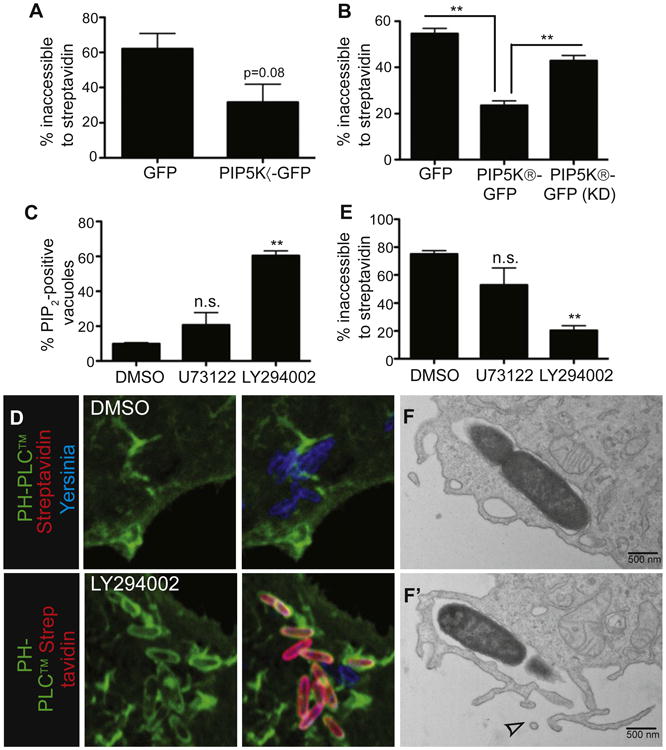
(A) Overexpression of PIP5Kα inhibits Yersinia entry into cells. COS cells transfected with the indicated constructs were infected with biotinylated Alexa 647-labeled Yersinia for 30 min, then stained with Alexa 546-streptavidin before fixation and quantification of bacterial entry.
(B) Overexpression of PIP5Kβ, but not a kinase-dead (KD) mutant, inhibits Yersinia entry into cells. Experiment conducted as in (A).
(C and D) Inhibition of PI3K increases the proportion of Yersinia at the PI(4,5)P2-containing stage. COS cells transfected with PH-PLCδ-GFP were treated with U73122, LY294002, or vehicle (DMSO), followed by infection with Alexa 647-labeled Yersinia for 30 min and fixation. PI(4,5) P2-positive bacteria were then quantified. (D) shows representative images from (C).
(E) PI3K inhibition reduces Yersinia entry into host cells. COS cells were treated with inhibitors, then infected with biotinylated Alexa 647-labeled Yersinia for 30 min, followed by staining with Alexa 546-streptavidin and fixation. Images were quantified to assess bacterial entry. Data are means of three (A and B) or four (C and E) independent experiments. Error bars represent ± SEM.
(F) Inhibition of PI3K blocks Yersinia entry just prior to scission. LY294002-treated COS cells were infected with Yersinia for 10 min, followed by fixation and processing for TEM. (F) and (F′) are serial sections; top section shows a seemingly sealed vacuole, which revealed an opening to the extracellular space in the subsequent section (F′, see arrowhead). See also Figure S2.