
Figure 7. The Final Stages of Yersinia Entry into Host Cells Involve PI3K-Dependent Fusion of Rab5-Positive Vesicles with Prevacuoles.
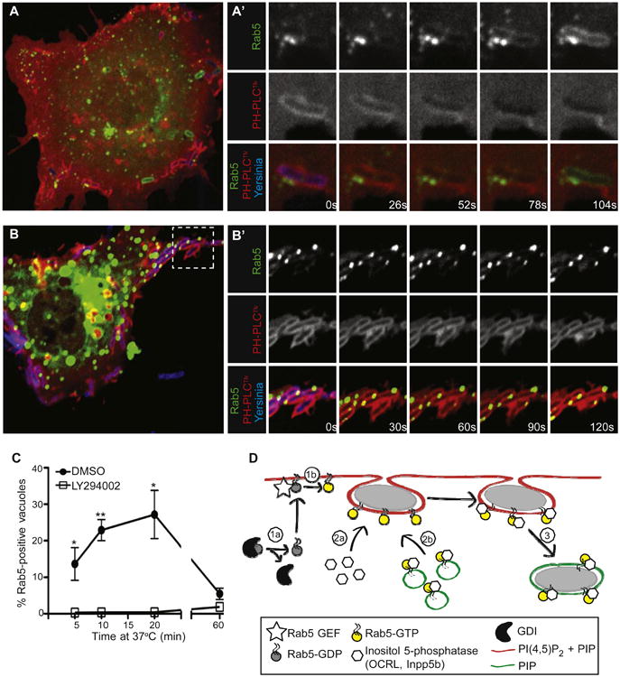
(A and A′) Rab5-positive vesicles are recruited to Yersinia-containing vacuoles seconds before PI(4,5)P2 is lost from their membranes. COS cells were cotransfected with Rab5-GFP and PH-PLCδ-RFP, and infected with Alexa 647-labeled Yersinia. Images in A′ were acquired at 26 s intervals.
(B and B′) Rab5-positive vesicles are recruited but cannot fuse with PI(4,5)P2-positive Yersinia-containing vacuoles in cells treated with LY294002. Experiment was conducted as in A, with images acquired every 30 s.
(C) COS cells were transfected with Rab5-GFP and treated with LY294002 or vehicle (DMSO) prior to infection for various times with Yersinia. Postinfection, cells were fixed, and Rab5-positive bacteria-containing vacuoles were quantified. Data are means of three independent experiments. Error bars represent ± SEM.
(D) Model of prevacuole scission. Rab5 may associate with the plasma membrane (1a), where it can be activated by plasma membrane-localized GEFs (1b), thereby allowing direct recruitment of OCRL or Inpp5b from the cytosol (2a). Alternatively (or additionally) endosomes bearing Rab5/OCRL/Inpp5b may fuse with the prevacuole membrane (2b). Recruitment of OCRL/Inpp5b to prevacuoles results in hydrolysis of PI(4,5)P2 and scission (3), generating a sealed vacuole. See also Figure S6 and Movie S1.