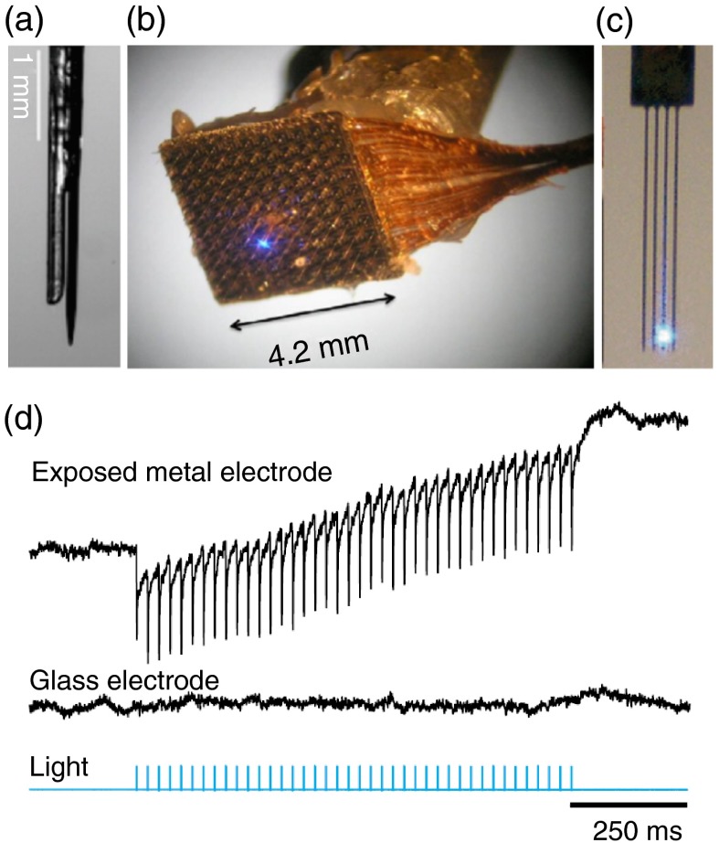
Fig. 3.
Optrode designs: (a) optrode design made from a core multimode fiber and a metal recording electrode;7 (b) integrated device that combines a multielectrode array and a fine illumination site;88 (c) multiarray silicon probes with integrated optical fibers for spatiotemporal brain control and recording;89 and (d) simultaneous local field potential recordings from mice tissue not expressing opsins with light-exposed metal (upper trace) and glass microelectrodes (middle trace). Light stimulation is represented in blue. Note the photo-electric artifacts in the upper trace (modified from Ref. 74).

