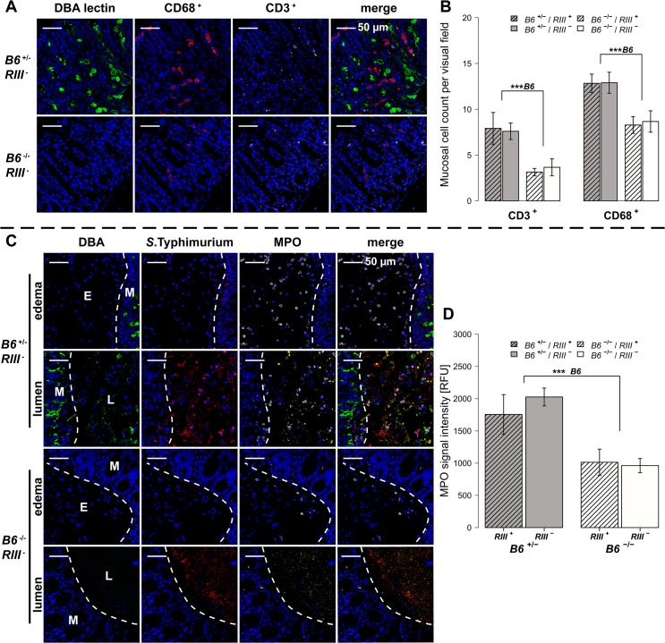
Fig 5. B4galnt2-dependent infiltration of immune cells after S. Typhimurium infection.
(A and B) Immunofluorescence staining and enumeration of positive cells per vision field showed that B6 +/- mice have higher numbers of CD68 (red) and CD3 (white) positive cells in the cecal mucosa 1d p.i. (N = 5–7). Nuclei were stained with DAPI (blue) and B4galnt2 glycans by using fluorescein labeled DBA (green). (C) Myeloperoxidase (MPO) positive cells (white) and S. Typhimurium (red) were determined by immunofluorescence staining. (D) MPO signal in lumen and edema was quantified and expressed as relative fluorescence units (RFU) (N = 7; Linear model; # P < 0.100, * P < 0.050, ** P < 0.010, *** P < 0.001, error bars indicate SEM).

