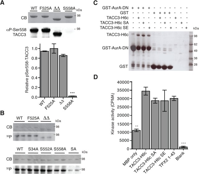
Fig 4. Biochemical characterisation of TACC3 mutants defective in either Aurora-A phosphorylation or activation.
(A) Ser558 phosphorylation of TACC3-H6c WT, F525A, ΔΔ (TACC3-H6c Δ519–546 and Δ564–629) and S558A by Aurora-A was measured by quantitative immunofluorescent blotting using an antibody specific to phosphorylated Ser558 in TACC3 (middle, the scanned blot was converted to black and white). The coomassie-stained gel is shown above. Quantification of phosphorylation is shown below. Error bars represent the standard error for two independent reactions. *** = P<0.001 using one-way ANOVA with Dunnett's post-hoc test compared to the WT reaction. (B) Total phosphorylation of TACC3-H6c WT, ΔΔ and point mutants by Aurora-A was monitored by incorporation of 32P-labelled ATP (bottom). Phospho-null (SA) has three mutations: S34A, S552A, S558A. Coomassie-stained gels are shown above. (C) Co-precipitation assay to assess binding between GST-AurA-DN and TACC3. The assay used wild-type, phospho-null (SA) and phospho-mimic (SE) TACC3-H6c as prey proteins. GST was used as a binding control. (D) Activation of Aurora-A by TACC3-H6c WT, SA and SE was monitored by in vitro kinase activity assay. The catalytic activity of Aurora-A was determined by incorporation of 32P into MBP and quantified by scintillation counting. TPX21-43 was used as a positive control for Aurora-A activation. Error bars represent the standard error for two independent reactions. ** = P<0.01 and *** = P<0.001 using one-way ANOVA with Dunnett's post-hoc test compared to the WT reaction.

