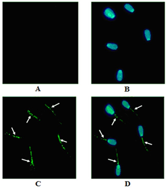
Fig 3. HIBADH is expressed and localized in bull spermatozoa.

The arrow heads indicate that HIBADH protein was mainly localized at sperm neck-piece and mid-piece, and to a lesser extent, in the sperm head. (A) Negative control. (B) Nuclear DNA signals (blue). (C) HIBADH protein signals (green). (D) Merging of HIBADH protein (green) and nuclear DNA signals (blue). Image was obtained using an inverted microscope (OLYMPUS) at 40 × 10. Images were obtained on a 5 μm scale plate.
