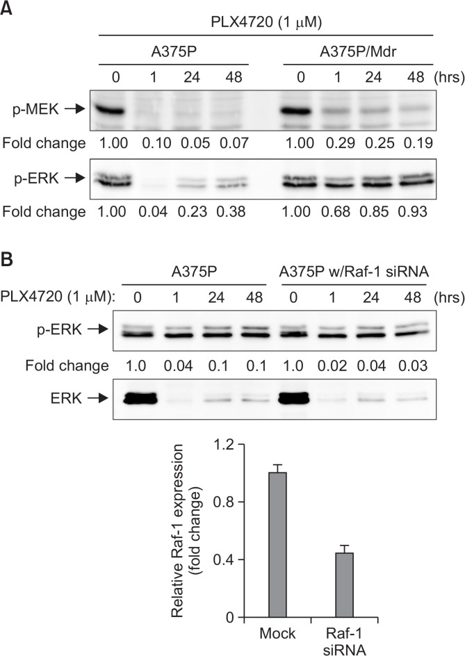
Fig. 4.
Rebound activation of pERK signaling after long-term treatment with PLX4720. (A) Cell lysates were prepared from BRAF inhibitor-sensitive (A375P) and BRAF inhibitor-resistant (A375P/Mdr) melanoma cells treated with PLX4720 (1 μM) for the indicated times. (B) BRAF inhibitor-sensitive A375P cells were transfected with siRNA pools targeting Raf-1 or scrambled oligonucleotides for 24 h. The cells were then treated with PLX4720 for the indicated times. (A, B) The phosphorylated forms of MEK and ERK were detected by immunoblotting using anti-phospho-MEK and anti-phospho-ERK antibodies. The same blots were reprobed with anti-MEK and anti-ERK antibodies to confirm that the expression levels of MEK and ERK proteins were similar in all of the lanes. Numbers listed below each band indicate the intensity value quantified by the Kodak Molecular Imaging software, expressed as fold change. The intensity value observed in vehicle control cells was defined as 1.0. The data are representative of at least two independent experiments. In lower inset of (B), siRNA-mediated knockdown of Raf-1 was confirmed by real-time RT-PCR analysis. The Raf-1 expression was normalized to the β-actin expression level. The relative expression level in mock control was regarded as 1.0.

