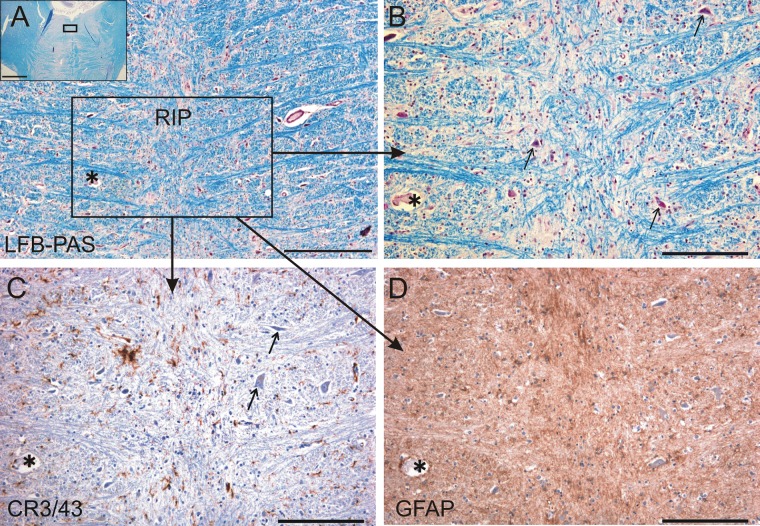
Fig 2. Histologically normal omnipause neurons.
Omnipause neurons in the nucleus raphe interpositus (RIP) appear histologically normal (arrows). (A) Photograph of a transverse section of the pons at the level of RIP. The inset indicates the area shown in A. (B) LFB-PAS staining showed no evidence of demyelination. (C) CR 3/43 staining showed no abnormal microglial activation. (D) GFAP staining showed no reactive gliosis. The asterisk labels the same blood vessel in neighboring sections for orientation. LFB-PAS, Luxol fast blue periodic acid-Schiff; GFAP, glial fibrillary acidic protein. Scale bar A = 500μm, inset = 5mm; B-D = 200μm.

