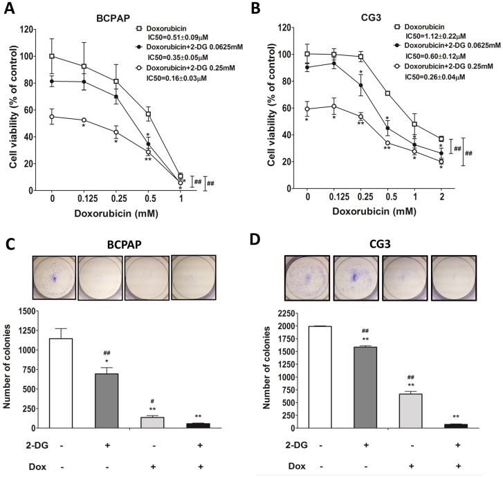
Fig 4. Effects of doxorubicin on the viability of PTC cells treated or not with 2-DG.
(A, B) BCPAP and CG3 cells cultured in medium supplemented with 10% fetal bovine serum were treated for 48 h with different concentrations of doxorubicin in the presence or absence of 2-DG. Post-treatment, cell viability was measured using a WST-1 assay. (C, D) Colony formation in BCPAP and CG3 cells treated for 48 h with 1 mM 2-DG, 0.5 μM doxorubicin, or both. The medium was replaced with fresh medium for 3–5 days to check for colony formation. *p < 0.05, **p < 0.001, compared to the controls (t test); panels A, B: ##p < 0.001, 2-way ANOVA; C, D: ##p < 0.001, compared to 2-DG plus doxorubicin treatment (t test). Data are presented as means± standard deviation (SD).

