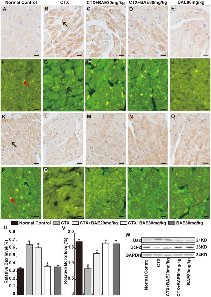
Fig 8. BAE attenuated CTX-induced cardiomyocyte apoptosis.
The heart sections were reacted with anti-Bax (A-J) or anti-Bcl-2 (K-T) monoclonal antibody(immunohistochemical staining and immunofluorescence, Bar: 20 μm). We observed that the Bax in CTX group was more intense than normal control group in cytoplasm, whereas Bcl-2 was highly expressed in myocardial cells in normal control group was more intense than CTX group. The black arrow represents respectively the Bax or Bcl-2-positive cardiomyocytes and the red arrow represents the nuclear of cardiomyocytes. Both doses of BAE significantly decreased Bax and increased Bcl-2 proteins expression in cardiac tissues. (U-W)Western blot was performed to detect the Bcl-2 and Bax proteins expression. All proteins were normalized to the corresponding GAPDH. Right: western blot image. Left: statistic data. #p < 0.05, compared to normal control; *p < 0.05, compared to CTX.

