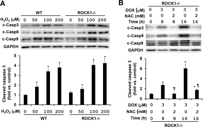
Fig 7. ROCK1 deletion has no inhibition on H2O2-induced caspase activation.
(A). Representative image of Western blot of cleaved caspases 3, 8, and 9 (top) and quantitative analysis (bottom) of Western blot of cleaved caspase 3 in cell lysates from attached WT and ROCK1 -/- MEFs treated with increasing concentrations of H2O2 for 4 h. Equal amount of proteins was loaded. (B) Representative image of Western blot of cleaved caspases 3, 8, and 9 (top) and quantitative analysis (bottom) of Western blot of cleaved caspase 3 in cell lysates from attached ROCK1 -/- MEFs treated with 3 μM doxorubicin and/or 2 mM NAC for 8 or 16 h. Similar experiments performed with the WT MEFs were presented in Fig 3B. n = 4–6 in each condition. * P < 0.05 vs. control of the same genotype. # P < 0.05 vs. WT under the same treatment condition. ¶ P < 0.05 vs. the same genotype under doxorubicin only condition.

