
Figure 1. Nonhuman primate venous thrombosis model and experimental design.
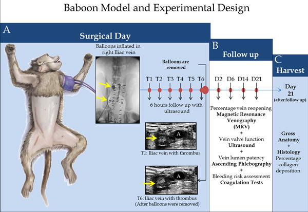
Schematic representation of the bi-balloon vein occlusion technique.
A) A balloon is placed and inflated in the iliac vein, just below the iliac bifurcation; another balloon is placed and inflated near the pelvic crest (yellow arrows); both balloons remain in initial position for 6 hours (T6) promoting thrombus formation. Baseline venogram; balloons inflated; hourly ultrasound follow-up; balloons removed; final ultrasound to confirm the presence of the thrombus at T6 and that a thrombus remains in a vein segment of approximately 3.5 cm in length. B) Percentage vein reopening magnetic resonance venography (MRV), Vein valve function ultrasound, vein lumen patency, ascending phlebography and bleeding risk assessment coagulation tests were evaluated at each time point: Days 2, 6, 14 and 21 after thrombosis. C) At day 21, after the follow up, a harvest of the tissue for gross anatomy and Histology (for leukocyte counts, quantification of percentage of thrombus fibrin and vein wall collagen deposition) were performed.