
Figure 5. Contrast Venography.
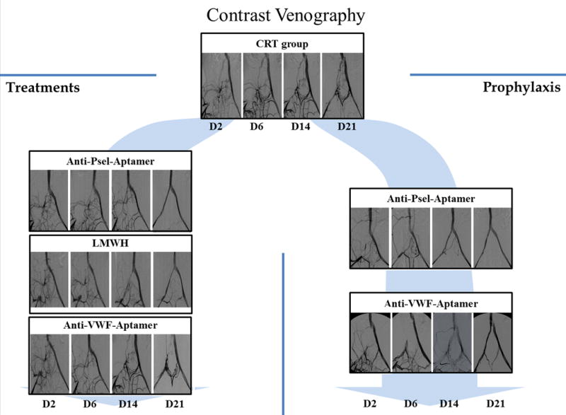
Selected pictures from contrast venography, performed at days 2, 6, 14 and 21 after thrombosis, that show the recanalization process. Occlusion of the iliac vein and collateral circulation was observed in all groups at day 2. Of note, recanalization was evident directly by the channel in the iliac vein and indirectly by the reduction of collateral circulation. CTR: control group; anti-Psel Aptamer-Px: P-selectin inhibitor prophylaxis group; anti-Psel-Aptamer-Tx: P-selectin inhibitor treatment group; LMWH-Tx: Low molecular weight heparin treatment group; anti-VWF-Aptamer-Px: von Willebrand factor inhibitor prophylaxis group; anti-VWF-Aptamer-Tx: von Willebrand factor inhibitor treatment group.