Figure 1.
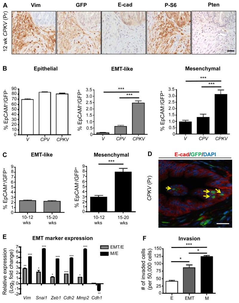
Tracking EMT and mesenchymal tumor cells in an endogenous prostate cancer model using a Vimentin-GFP reporter line. A, Prostates from CPKV mice (12 weeks) display EMT regions that are Vimentin (Vim)/GFP-positive surrounding E-cadherin (E-cad)-positive epithelial glandular structures. P-S6 marks cells that have undergone Cre recombination. B, Vimentin-GFP (GFP) and EpCAM were used to characterize epithelial (EpCAM+/GFP−), EMT (EpCAM+/GFP+), and mesenchymal (EpCAM−/GFP+) cell populations from the prostates of various Vim-GFP reporter mice (10–12 weeks) by FACS. CPKV mice have significant expansion of EMT and mesenchymal tumor cell populations. C, Expansion of mesenchymal tumor cell populations in CPKV prostates during late stage tumor progression (15–20 weeks). D, EMT cells (yellow arrow) that are GFP+ (green) and E-cadherin (E-cad)+ (red) are found within epithelial glandular structures in CPKV prostates (12 weeks). E, qPCR analysis confirms EMT signature gene expression in EMT and mesenchymal tumor cells isolated from CPKV prostates (10–12 weeks). F, Matrigel invasion assay reveals that EMT and mesenchymal tumor cells isolated from CPKV prostates (10–12 weeks) are significantly more invasive than epithelial tumor cells. Data in B, C, E, and F are represented as mean ± SEM. Bar, 50 μm. Pr, prostate. Lin−,CD45−/CD31−/Ter119−. *, P < 0.05, **, P < 0.01. ***, P < 0.001.

