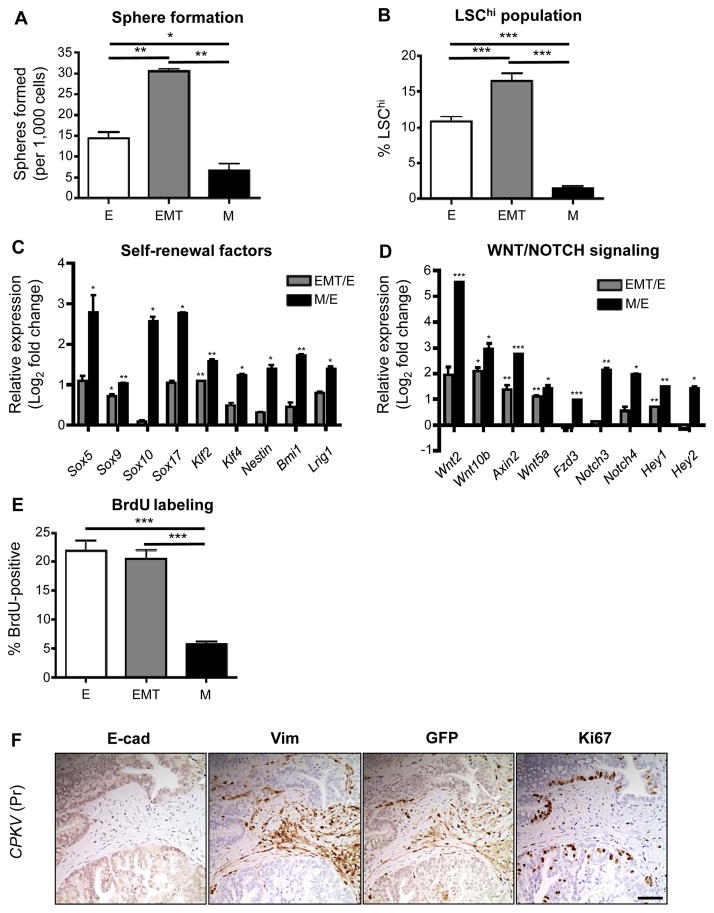
Figure 2.
EMT and mesenchymal tumor cells have enhanced stemness properties. A, Matrigel sphere assay reveals that EMT tumor cells sorted from CPKV prostates (10–12 weeks) form more spheres than epithelial and mesenchymal tumor cells after 7 days in culture. B, EMT tumor cells have a higher LSChi content compared to epithelial and mesenchymal tumor cells from CPKV prostates (10–12 weeks), as quantified by FACS. C, EMT and mesenchymal tumor cells from CPKV prostates (10–12 weeks) have enhanced expression of self-renewal and stemness factors compared to epithelial cells. D, EMT and mesenchymal tumor cells from CPKV prostates (10–12 weeks) have enhanced expression of genes involved in WNT and NOTCH signaling compared to epithelial cells. E, Epithelial and EMT tumor cells from CPKV prostates (10–12 weeks) have a higher percentage of cells in S-phase compared to mesenchymal tumor cells, as measured by the percentage of BrdU+ cells. F, Ki67-positive cells are found preferentially in E-cadherin (E-cad)-positive glandular structures compared to Vimentin (Vim)/GFP-positive EMT regions in the stroma of CPKV prostates (12 weeks). Data in A through E are represented as mean ± SEM. Bar, 100 μm. Pr, prostate. *, P < 0.05, **, P < 0.01. ***, P < 0.001.

