Figure 4.
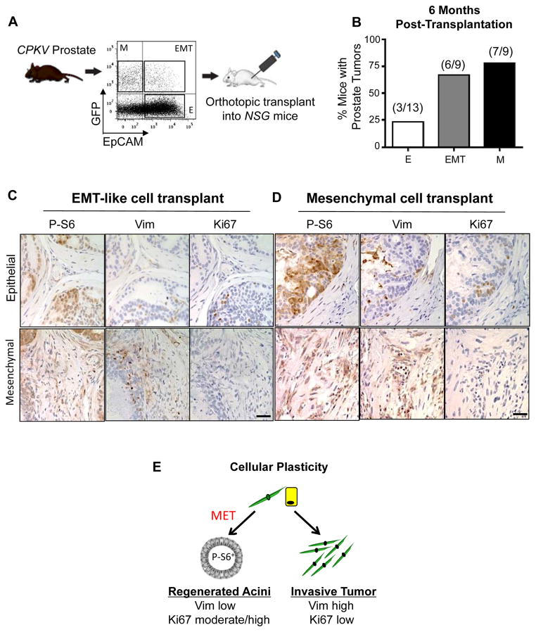
EMT and mesenchymal tumor cells have enhanced tumor-initiating capacity and cellular plasticity in vivo. A, Schematic of experimental design for orthotopic transplantations. B, EMT and mesenchymal tumor cells form tumors more readily in vivo compared to epithelial tumor cells from CPKV prostates (10–12 weeks). C, IHC stains of anterior lobes from an NSG mouse transplanted with EMT tumor cells from CPKV prostates (10–12 weeks). P-S6 was used to trace transplanted cells, and Vim was used to mark mesenchymal cells. Top panel, transplanted EMT tumor cells form regenerated glandular structures. Bottom panel, transplanted EMT tumor cells form invasive mesenchymal tumor regions. D, Same as in C, except a prostate section from an NSG mouse transplanted with mesenchymal tumor cells from CPKV prostates (10–12 weeks). E, EMT (yellow) and mesenchymal (green) tumor cells from CPKV prostates have the plasticity to undergo an MET and regenerate epithelial glandular structures, or form invasive mesenchymal tumors in vivo. Bar, 50 μm.

