Figure 6.
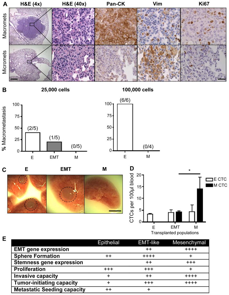
Epithelial tumor cells have enhanced metastatic seeding potential. A, IHC analysis of epithelial (Pan-CK), mesenchymal (Vim), and proliferation (Ki67) markers in micrometastases (micromets) and macrometastases (macromets) in primary CPKV lungs (18 weeks). Low magnification bar, 500 μm; high magnification bar, 50 μm. B, Percent of macrometastatic lesions in the lungs of NSG mice 16 weeks after intravenous transplantation of either 25,000 or 100,000 epithelial, EMT, or mesenchymal tumor cells from CPKV prostates (10–12 weeks). C, Whole-mount images of lungs isolated from NSG mice transplanted with 25,000 epithelial, EMT, or mesenchymal tumor cells from CPKV prostates (10–12 weeks). Circle, macrometastases. Bar, 4 mm. D, NSG mice transplanted intravenously with mesenchymal tumor cells (25,000) from CPKV prostates (10–12 weeks) contained a significantly higher number of CTCs persisting in the bloodstream compared to mice transplanted with either epithelial or EMT tumor cells 16 weeks post-transplantation. E, Table summarizing the characteristics of epithelial, EMT, and mesenchymal tumor cells isolated from CPKV prostates. Data in D are represented as mean ± SEM. *, P < 0.05

