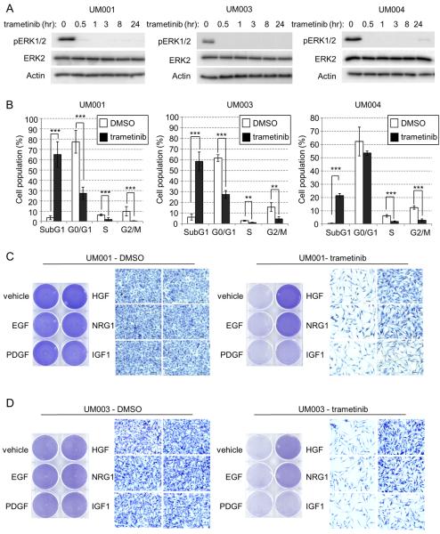
Figure 1. NRG1 and HGF rescue growth abrogation induced by MEK inhibitors in UM cells.
(A) UM001, UM003 and UM004 cells were treated with 100 nM of trametinib (GSK1120212) for the indicated times. Cell lysates were probed with phospho ERK1/2, total ERK2 and actin antibodies. (B) UM001, UM003 and UM004 cells were treated with DMSO or trametinib for 3 days (UM001 and UM004 cells) or 5 days (UM003). Cells were then fixed, permeabilized and subjected to propidium iodide (PI) staining. Cell cycle analysis was performed with FlowJ software. *P<0.05, **P<0.01, ***P<0.001, based on two-tail Student's t-test assuming unequal variance. (C) UM001 cells were treated with vehicle control, 10 ng/ml of EGF, PDGF-B, HGF, NRG1 and IGF1 alone or together with 100 nM trametinib. After 72 hr, cells were subjected to crystal violet staining. Representative microscopic images of the cells at 200× magnification are shown. Scale bar is equal to 50 μm. (D) UM003 cells were treated as in C for a total of 5 days. Drugs and growth factors were replenished on day 3. Cells were stained with crystal violet.

