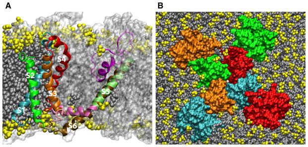
Fig. 2.

The structure of the KV1.2 paddle-chimera channel (Long et al. 2007) in a lipid bilayer. a Cut-away view highlighting a single protein chain colored by transmembrane segment (S1, cyan; S2, green; S3, orange; S4, red; S5 lime; S6, ochre). The connecting region between S1 and S2 is not shown for clarity. The other three protein chains are shown in a transparent molecular surface representation colored white. Lipids are shown as filled-spheres with the headgroups in yellow. b Top view. The pore domain results from the assembly of the S5 through S6 regions of the four protein chains. The four S1–S4 VSDs are on the periphery of the pore domain. The protein chains are shown in a molecular surface representation. Lipids are shown as filled-spheres (headgroups in yellow) and K+ ions are shown as white spheres. The images correspond to a configuration from an all-atom molecular dynamics simulation of the full-length KV1.2 paddle-chimera channel embedded in a lipid bilayer in excess water (the latter are not shown for clarity) (Color figure online)
