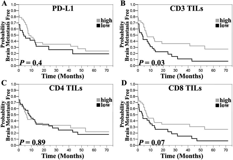
Figure 4.
Kaplan-Meier curves showing the association between PD-L1 expression or TIL density and time to development of brain metastases from the time of diagnosis of metastatic melanoma. The median PD-L1 intensity score was utilized to dichotomize scores into low/high while the median CD3 TIL areas was used as thresholds for defining high/low TIL density. High density of CD3 positive TILs was significantly associated with a longer time to development of brain metastasis.

