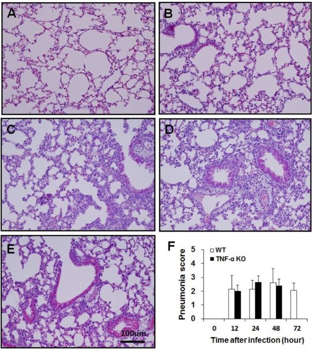
Figure 3. Histopathological analysis of lungs from WT and TNF-α KO mice after intranasal S. pneumoniae infection. The lungs of WT (A, C, E) and TNF-α KO (B, D) mice at 0 (A, B), 48 (C, D), and 72 h (E) post-infection. Sections were stained with H&E. The degree of pneumonia was scored and expressed as the means±SD (F).