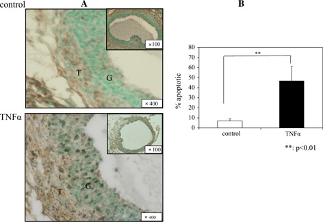
Figure 5.

TUNEL staining 12 h after hCG treatment and rate of apoptosis. Apoptotic cells were detected by TUNEL‐positive cells (brown staining). Many apoptotic cells were seen in the granulosa of the TNFα group compared with the control group (a). The rate of apoptotic nuclei was significantly higher in the TNFα group than in the control group (p < 0.01) (b). G granulosa cell, T theca cell
