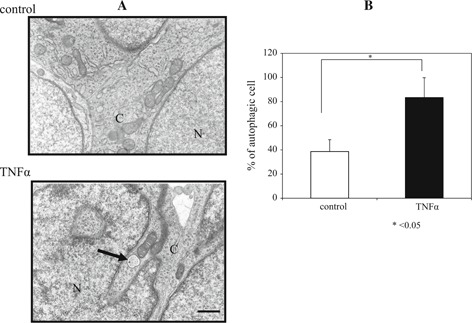
Figure 6.

Transmission electron microscopic image of granulosa cells 12 h after hCG treatment. Autophagic vacuoles were present in the cytoplasm in the TNFα group (arrow) (a). The rate of autophagic vacuoles was significantly higher in the TNFα group than in the control group (p < 0.05) (b). N nucleus, C cytoplasm, bar 500 nm
