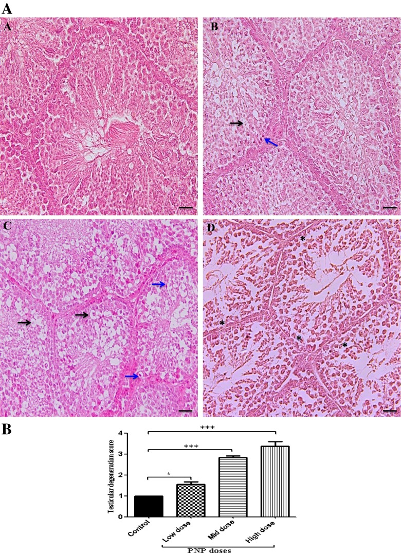
Fig. 5.
Microscopic examination (H&E stain) of the testes in the control sections (a) showed normal architecture and clear compartmentalization in the seminiferous tubules, with spermatozoa visible in normal-sized lumen. The PNP-treated groups; the low dose (b), the mid dose (d), and the high dose (e) showed vacuolation of some spermatogenic cell and spermatocyte (black arrows), karyopyknosis (blue arrows), and hypocellularity of spermatogenic cell and spermatocyte (asterisks), as well as cellular degeneration. The scale bar represents 50 μm. b Quantitative analysis of testicular degeneration rates in the testes of the control and the PNP-treated quails. Data are expressed as average of the scores ± S.E.M. in 250 seminiferous tubules/animal (n = 4). The degeneration scores ranged from normal (1) to mild (2), moderate (3), and severe (4). ***P < 0.001 in the PNP-treated groups vs. the control group

