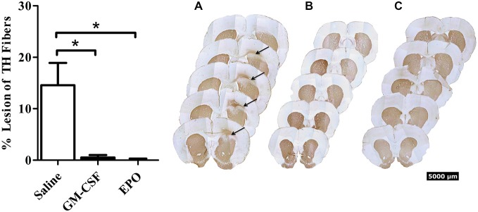
Figure 1.

Thirty days after the intra-striatal infusion of 6-OHDA, saline treated animals (graph—white bars; A) had a modest but statistically significant loss of TH+ striatal fibers. The GM-CSF (graph—black bars; B) and EPO (graph—black bars; C) treated animals displayed no visible lesion at the 30-day sacrifice time. Data is expressed as mean ± 1 SEM, *p < 0.01.
