. 2015 Jun 26;18(2):200–205. doi: 10.4048/jbc.2015.18.2.200

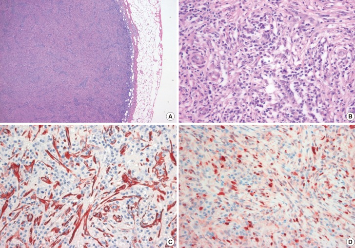
Figure 2. Histologic, immunohistochemical analyses of inflammatory myofibroblastic tumor (IMT) at initial diagnosis. (A) A sample of the IMT obtained on excisional biopsy shows mainly composed of spindle cells forming swirling storiform-like patterns, as well as inflammatory cells including plasma cells and lymphocytes (H&E stain, ×100). (B) The mass showed high mitotic activity and mild cellular pleomorphism (H&E stain, ×400). (C) Immunohistochemical analysis shows positive staining for smooth muscle actin (SMA) in the spindle tumor cells (SMA, ×400) and (D) positive staining for Ki-67 in about 30% of the spindle tumor cells (Ki-67, ×400).

Figure 2