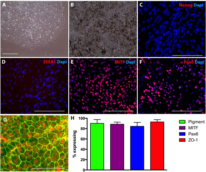
Figure 1. Generation of porcine iPSC derived RPE cells.
A: Representative phase micrograph of pig iPSC colonies consisting of densely packed cells with high nucleus to cytoplasm ratio. B: Densely pigmented porcine iPSC-derived RPE cells with typical hexagonal morphology. C–G: Immunocytochemical analysis of porcine iPSC derived RPE cells targeted against Nanog (C), SSEA1 (D), MITF (E), Pax6 (F & G) and ZO-1 (G). H: Histogram depicting the total number of cells post-differentiation that were pigmented, expressed MITF, Pax6, and ZO-1.

