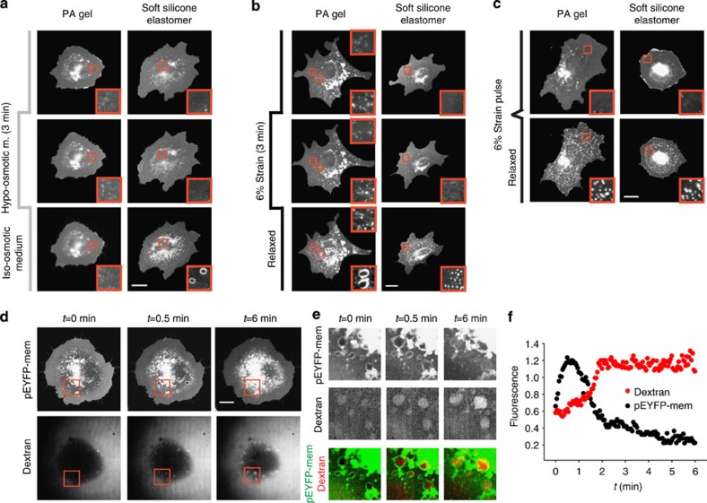
Figure 3. VLD formation is driven by the confinement of liquid flows at the cell–substrate interface.
Response of pEYFP-mem-transfected cells seeded on either poly-acrylamide (PA) gels or soft silicone elastomers to: (a) the application of 50% hypo-osmotic medium for 3 min, (b) the application of 6% strain for 3 min and (c) a fast 6% strain pulse. Insets show zoomed views (10x10 μm2) of membrane structures. Scale bars, 20 μm. No significant differences were observed between any of the cases either in the diameter of reservoirs (n=150 reservoirs from 3 cells) or in their density (n=30 cell regions from 3 cells). (d) Time sequence of VLD formation and resorption in pEYFP-mem-transfected cells exposed to dextran-labelled iso-osmotic media after 3 min incubation with 50% unlabelled hypo-osmotic media. (e) Zoomed insets (20 × 20 μm2) corresponding to red square in d showing the evolution of membrane and dextran fluorescence, and merged images. (f) Corresponding quantification of pEYFP-mem and dextran relative fluorescence levels.

