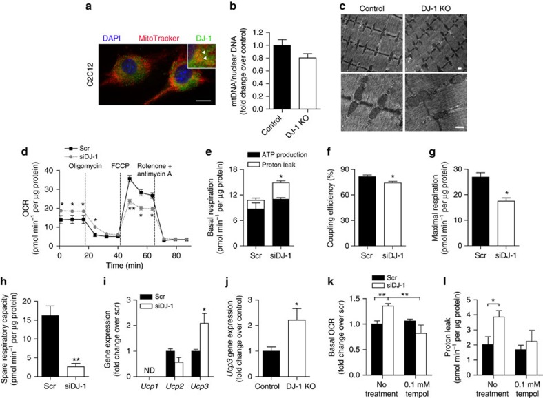
Figure 4. DJ-1 deficiency induces muscle mitochondrial uncoupling via ROS.
(a) Representative micrograph of C2C12 myoblasts stained with MitoTracker and antibody against DJ-1. Scale bar, 10 μm. Inset: higher magnification image. Arrowhead, co-localization of DJ-1 with MitoTracker. (b) mtDNA copy number calculated as the ratio of Cox2 to Ppia levels measured by real-time quantitative PCR in quadriceps tissue. Female mice were fed a HFD for 3 months starting at 2 months of age (n=5 per group). (c) Transmission electron microscopy images from quadriceps tissue of HFD-fed female mice. Scale bar, 500 nm. (d) OCR in C2C12 myotubes measured using the Seahorse flux analyzer in response to 1 μM rotenone, 0.5 μM FCCP, and 1 μM rotenone and antimycin A (n=3 per group). Experiments were repeated at least three times. (e–h) ATP production, proton leak, coupling efficiency, maximal respiration and spare respiratory capacity calculated from (d). (i,j) mRNA expression of uncoupling proteins measured by quantitative RT–PCR in (i) C2C12 myotubes (n=3–6 per group) and (j) quadriceps tissue of HFD-fed female mice (n=6 per group). ND, not detected. (k) Basal OCR and (l) proton leak in myotubes treated with 0.1 mM tempol (n=3 per group). Results are presented as mean±s.e.m. according to the two-tailed unpaired Student's t-test. *P<0.05; **P<0.01.

