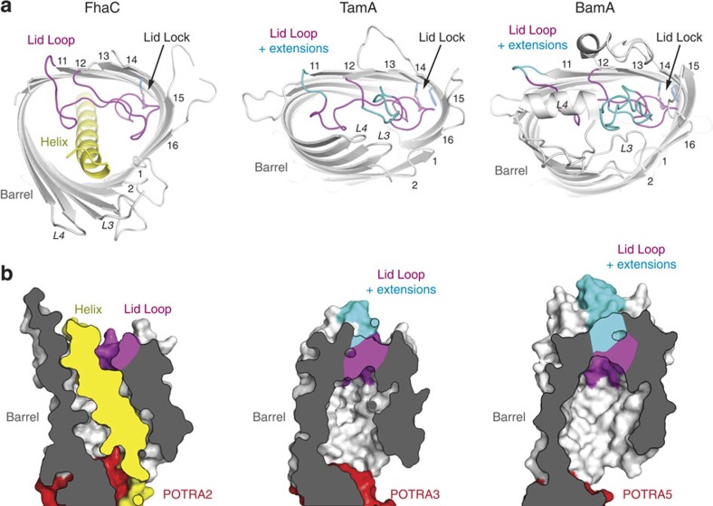
Figure 5. Barrel shape, lid lock and H1 helix insertion in FhaC.
(a) Top-view ribbon representation of the barrels of B. pertussis FhaC (left; PDB entry 4QL0; this work), E. coli TamA (center; PDB entry 4C0012) and H. ducreyi BamA (right; PDB entry 4K3C14). The β-barrels are shown grey, the helix of FhaC is shown yellow. Using the same colour code as in Fig. 4, conserved regions of loop L6 are shown in magenta, extensions of L6 in TamA and BamA relative to FhaC are shown in cyan. (b) Cross-sectional surface representation of the barrels of B. pertussis FhaC (left), E. coli TamA (center) and H. ducreyi BamA (right), in the same colour code. POTRA domains are shown in red.

