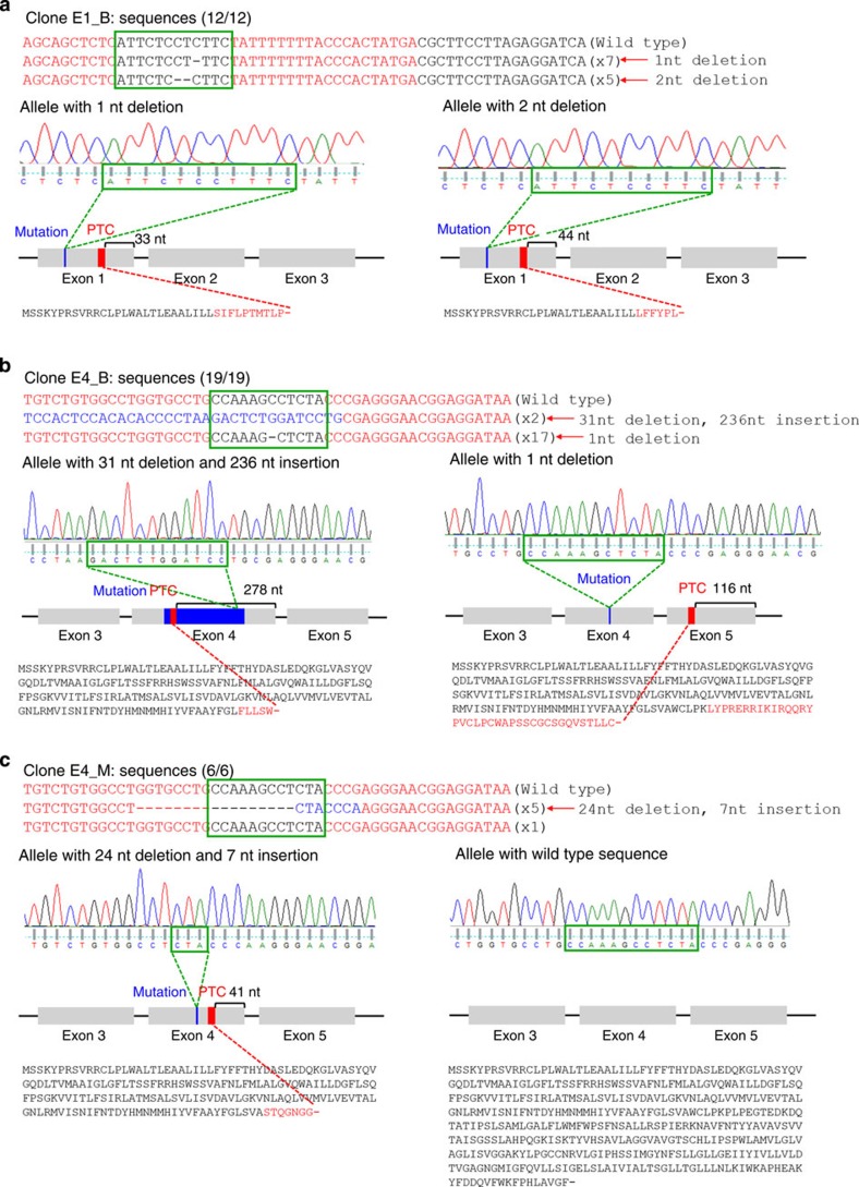
Figure 3. DNA sequences of RHD-mutated clones.
The RHD gene DNA sequences from the parental cells, clones with biallelic mutations in exon 1 (E1_B; a) or exon 4 (E4_B; b), and a clone with a monoallelic mutation in exon 4 (E4_M; c). TALE-binding sites are in a red font and spacer regions are indicated with green boxes. Deleted bases are indicated by dashes and inserted bases are shown in a blue font. The number of occurrences is shown in parentheses (for example, × 7 and × 5 indicate the number of each sequence). The sequence and sequencing chromatogram for each allele are shown. The locus of each mutation, the PTC generated by the mutation and the distance between the PTC and the exon–intron junction are depicted in a schematic of the RHD gene. Expected protein sequence translated from each allele are displayed, such that mutated protein sequences generated by a nuclease-induced frameshifting mutation are shown in a red font and translation termination is indicated with a dash. nt, nucleotide.

