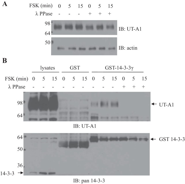
Fig. 3.
Dephosphorylation of UT-A1 by protein phosphatase 1. UT-A1 MDCK cells treated with 10 μM FSK for 0, 5, and 15 min were lysed with 1% Nonidet P-40 buffer. Cell lysates were incubated with or without lambda protein phosphatase (λ PPase) at 30°C for 30 min. Next, cell lysates were used for Western blot analysis for UT-A1 expression (4–20% SDS-PAGE gradient gel; A) or applied for GST-14-3-3 pulldown assay (10% SDS-PAGE gel; B).

