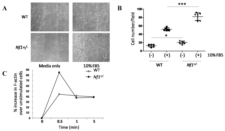
Figure 2.
Migration and actin polymerization were significantly enhanced in Nf1+/− MSPCs. (A) Wound healing assays were performed by incubating WT and Nf1+/− MSPCs in 10 µg/mL of mitomycin C for one hour, after which a linear wound (marked by the white dotted lines) was created as shown. Wound healing was allowed to proceed in fresh media for 24 h, (original magnification ×200); (B) The number of cells migrating into the wound field were quantified, revealing an increased migration in Nf1+/− MSPCs compared with WT MSPCs (F = 75.76, Df = 1, *** p < 0.001; *** p < 0.001 for Nf1+/− MSPCs vs. WT MSPCs in the presence of 10% FBS, * p < 0.05 for untreated Nf1+/− MSPCs vs. untreated WT MSPCs). Data are represented as mean ± SD from duplicate wells from three independent experiments, each experiment was performed with different MSPCs culture isolated from individual mice; (C) Actin polymerization was measured following 2 h starvation and subsequent treatment with 10% FBS for different time periods. Flow cytometry analysis was performed following 400 nM FITC-phalloidin staining. An increased F-actin content was observed in Nf1+/− MSPCs comparison to WT MSPCs. A representative result of one of three independent experiments is shown; each experiment was performed with different MSPCs culture isolated from individual mice.

