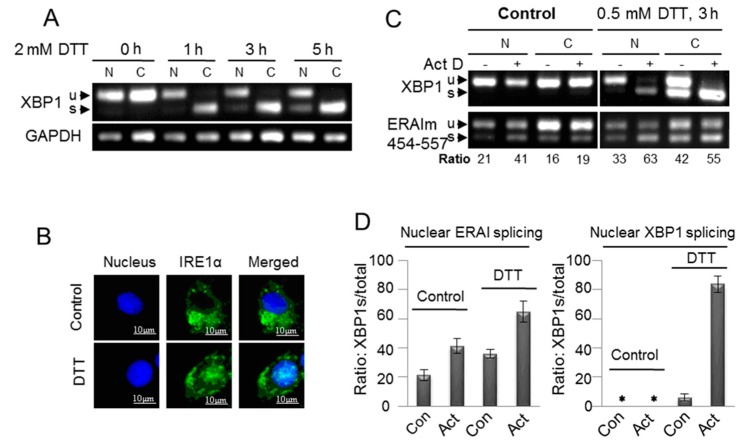
Figure 5.
Promotion of the unconventional splicing of XBP1 mRNA in the nucleus by acute ER stress. (A) MCF-7/wt cells were fractioned after they were treated with 2 mM DTT for different time as indicated. The fractioned RNA was subjected to RT-PCR with XBP1 primers; (B) MCF-7/wt cells were treated with 2 mM DTT for 5 h, and then were fixed. The immunoflurorescence of the fixed cells stained with monoclonal IRE1α antibody was imaged, as described in the Methods; (C) MCF-7/ERAIm454–557 cells were fractioned after they were treated with 0.5 mM DTT and/or 10 μg/mLK Actinomycin D (Act D) (pretreatment for 1 h) or vehicle for 3 h. The ratio shows the percentage of spliced mRNA in total ERAIm454–557 mRNA including spliced and unspliced mRNA; and (D) Quantified fractions of nuclear spliced XBP1 or ERAI mRNA, the ratio of spliced XBP1 mRNA to total XBP1 mRNA. Data are from three independent experiments as described in (C). Error bars show means ± SD. * means there was no stripe.

